Right left superior and inferior. The mediastinal surface faces the midline.
Physical Therapy For Cardiovascular Disorders Ppt Download
Houses vital centers for control of the heart respiration and blood pressure 6.
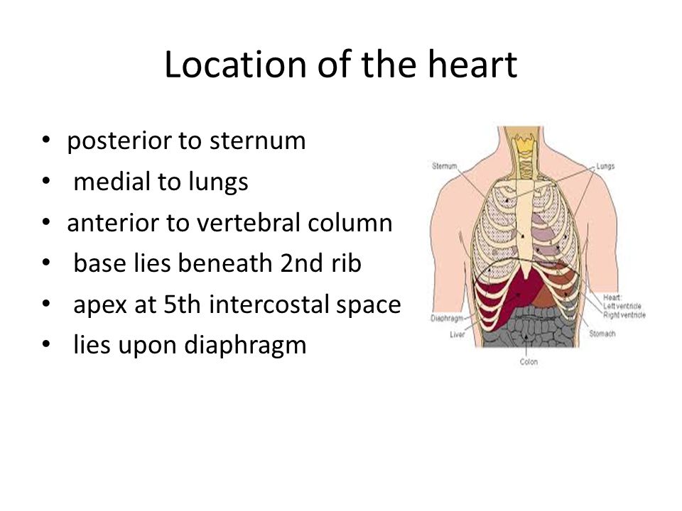
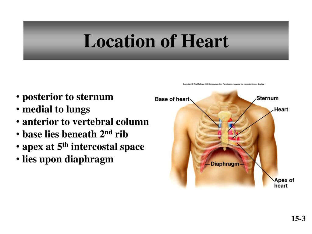
. The human heart is located within the thoracic cavity medially between the lungs in the space known as the mediastinum. The urinary bladder is __ to the uterus. A base is resting on the diaphragm.
The heart is situated in the middle of the two lungs and in front of a vertebral column in a thoracic cavity. Endocardium the thin inner lining of the heart chambers that also forms the surface of the valves. The posterior border is smooth and rounded in contrast to the anterior and inferior borders which are sharp.
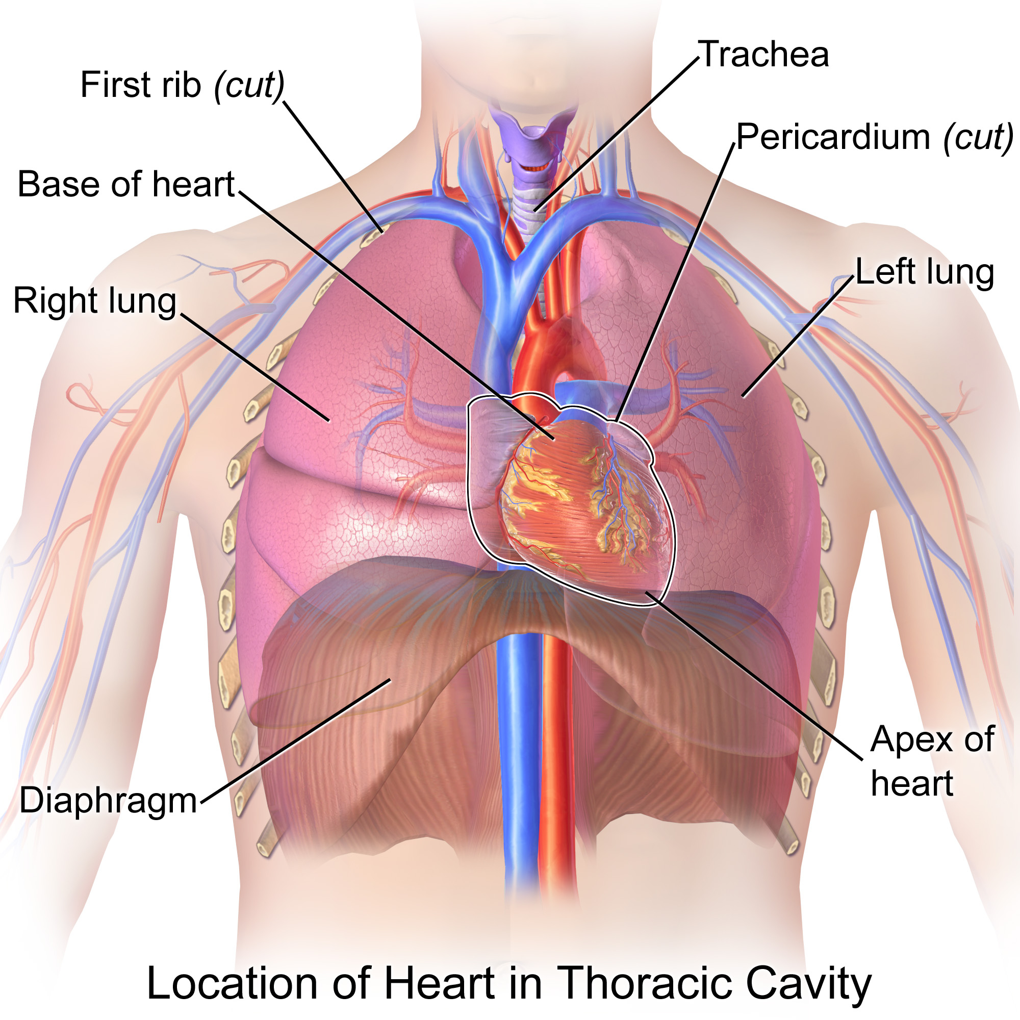
Its size is about that of a fist and its weight is about 250-300 g. The cardiac notch is a concavity in the lung that formes to accommodate the heart. Figure 1 shows the position of the heart within the thoracic cavity.
The inferior border separates the base of the lung from the costal and mediastinal surfaces. What side is your heart on. The heart is situated in the middle of the two lungs and in front of a vertebral column in a thoracic cavity.
Make sure you know the basics of lung cancer including prevention risk factors symptoms and. Both of the anatomy of the lungs are conical in shape and has the following features. The gallbladder is on the _____ surface of the liver.
It pumps blood throughout the body and is located behind the breastbone between the lungs. The cardiac notch is an indentation on the surface of the left lung and it allows space for the heart Figure 2221. The heart is located in the chest between the lungs behind the sternum and above the diaphragm.
The heart and lungs together with the blood vessels comprise the cardiovascular system. Figure 2221 Gross Anatomy. There is a rounded posterior border that separates the vertebral part of the medial surface from the costal part.
Your ribcage protects your heart. During the intrauterine stage the septum between the two atrial is open and a ductus connects the pulmonary artery to the aorta effectively bypassing the pulmonary circulation because the lungs are not functional. The heart has five surfaces.
The heart sits in the mid chest extending into the left side Oxygen is inhaled and released from the lungs to the blood. The stomach is _____ to the spleen. Each lung is located near different organs in the body.
Is the heart connected to the lungs. The lung extends from the ribs in front to the ribs behind and from the dome of the pleural cavity down to the diaphragm. It is known as the cardiac notch.
A large part of each lung lies behind the heart. The left lung lies close to the heart thoracic aorta and esophagus while the right lung is by the esophagus heart both vena cavas inferior and superior and the azygos vein. Two surfaces of the lungs are costal and medial.
Anatomy Where is your heart located. Your lungs as you probably know are a pair of highly elastic and spongy organs that sit inside your chest on either side of your heart. The posterior vertebral part can be found next to the thoracic vertebra and their associated intervertebral discs.
It also has several margins. The human heart is located within the thoracic cavity medially between the lungs in the space known as the mediastinum. Air reaches the alveoli air sacs where oxygen then moves from the.
The apex of the lung is the superior region whereas the base is the opposite region near the diaphragm. The pumped blood carries oxygen and nutrients to the body while carrying metabolic waste such as carbon dioxide to the lungs. Your heart is slightly on the left side of your body.
Features of the lungs. It is formed by the costal and. Its center is located about 15 cm to the left of the midsagittal plane.
It then returns the blood to the heart which pumps the freshly oxygenated blood to the rest of the body. However it is located posterior behind to the breastbone plate ie sternum. In order to understand how that happens it is necessary to understand the anatomy and physiology of the heart.
The heart is bounded laterally by the lungs in the posterior by the esophagus and trachea and in the anterior by the rib cage. The costal surface of the lung borders the ribs. On the left lung the anterior border is marked by a deep notch created by the apex of the heart.
The heart pumps blood from the body to the lungs where the blood is oxygenated. An apex at the upper end. The heart is medial to the lungs.
The heart is a strong and muscular cone-shaped organ that is about the size of a fist. The right margin is the small section of the right atrium that extends between the superior and inferior vena cava. Deoxygenated blood flows from the heart to the lungs where it gives up wastes and is freshly oxygenated.
Hereof are the lungs in front or behind the heart. However it is located posteriorbehind to the breastbone plate ie sternum. The inferior border is thin and separates the base of the lung from the costal surface.
The lungs are _____ to the heart. The heart and lungs are located in the thorax or chest cavity. It sits between your right and left lungs.
Brain area through which all the sensory input is relayed to get to the cerebral cortex 7. It is surrounded by the pericardium. Unlike other lung borders the posterior border is an imaginary line that coincides with the heads of the adjacent ribs.
It sits slightly behind and to the left of your sternum breastbone. The kidneys are _____ to the small intestine. However it is located posteriorbehind to the breastbone plate ie sternum.
The left lung is slightly smaller to make room for the heart in your left chest. Your heart is located in the front of your chest. The space between the two.
The heart develops from two endocardial tubes that merge loop and septate to form the heart. Brain area most concerned with equilibrium body posture and coordination of motor activity. The posterior border is thick and extends from the C7 to the T10 vertebra which is also from the apex of the lung to the inferior border.
Base posterior diaphragmatic inferior sternocostal anterior and left and right pulmonary surfaces. Is The Heart Located Posterior Or Medial To The Lungs Socratic Lung Anatomy Physiopedia. Location of the Heart.
Three borders of the lungs are anterior posterior and inferior borders.
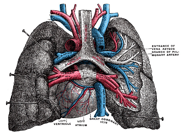
Figure Posterior View Of Heart And Statpearls Ncbi Bookshelf
Is The Heart Located Posterior Or Medial To The Lungs Socratic

Human Heart Lungs With Diaphragm Zoom With Anatomy Posterior View Stock Photo Picture And Royalty Free Image Image 85748670
Posterior View Of Heart And Lungs Stock Photo Alamy

Heart Flashcards Quizlet

Lungs And Structures Within Mediastinum Posterior View Diagram Quizlet

Lungs And Heart Posterior View Labelled Illustration Stock Image C043 4864 Science Photo Library
Chapter 15 Cardiovascular System Ppt Download
0 comments
Post a Comment